.webp)
Veterinary Pathology: Animal Health & Diagnostics
Rubbing your Golden Retriever’s belly usually feels routine, until you notice a new lump. Your local vet can examine what is visible from the outside, but they still need testing to understand what is happening inside. Clinical guidance often favors prompt testing over a wait-and-see approach because delay can put the animal at greater risk.
Much of that work happens behind the scenes. A veterinary pathologist studies cells, tissues, and fluids to identify disease. To them, a small tissue sample is not just a slide. It is the evidence that helps explain what is wrong.
The lab report does more than list results. It helps guide the next step in care. Veterinary pathology gives clinics clear answers so treatment plans are based on evidence, not guesswork.

Scispot for Veterinary Pathology and Clinical Diagnostics
In veterinary pathology and clinical diagnostics, Scispot helps labs move faster without losing track of their data. It gives teams one place to manage samples, test workflows, pathology records, instrument data, and reports, so information does not end up split across spreadsheets, paper notes, or disconnected systems.
From cytology and histopathology tracking to bloodwork, biopsy intake, result review, and final report readiness, Scispot helps veterinary labs run a more organized and traceable process. For labs that want clearer workflows, better visibility, and a system that can grow with testing volume, Scispot is a strong digital option.
From Blood Drops to Biopsies: Decoding the Two Main Branches of Animal Pathology
When your Golden Retriever seems unusually tired, the first clue may come from a routine blood draw. That sample falls under veterinary clinical pathology. This branch focuses on fluids like blood and urine to detect internal problems that are not obvious during a physical exam.
Sometimes the issue is a physical change, like a lump under your cat’s skin. That is where anatomic pathology comes in. Specialists examine solid tissues, often through histology, which looks at tissue structure under the microscope and shows how cells are arranged inside the mass.
Together, these fields form the backbone of modern veterinary diagnostics. Looking at anatomic and clinical pathology side by side helps specialists separate problems that may look similar at first glance, such as a fluid-filled infection and a solid tumor.
Sometimes vets can get useful answers with only a small group of cells, without moving straight to a surgical biopsy. That is where fine needle aspirates come in.
The Needle and the Microscope: Why Your Vet Performs Fine Needle Aspirates
Finding a new bump on your dog can be alarming. Still, getting answers does not always mean surgery. A fine needle aspirate lets a vet sample cells from a lump with a thin needle, usually in seconds, and gives a quick, non-surgical look at what may be going on.
This method depends on cytology, which is the study of individual cells. By examining those cells on a slide, pathologists can often screen for cancer and other problems without making a large incision.
That early look can give pet owners either reassurance or a clearer next step. Common findings in an FNA report include:
- Lipomas: benign clusters of normal fat cells
- Inflammation: immune cells responding to infection or irritation
- Abnormal growths: suspicious cells that may lead to further discussion around oncology and tumor grading
Sometimes, though, individual cells do not tell the full story. In those cases, pathologists need to examine the full tissue structure.
Reading the “Blueprint” of Disease: How Microscopic Tissue Samples Reveal the Truth
Sometimes a scattered group of cells is not enough. When vets need to see how a disease behaves in the tissue itself, they turn to histopathology. This involves studying an intact piece of tissue under the microscope.
To do that, the lab needs a biopsy. That tissue sample shows how abnormal areas relate to the healthy tissue around them, which is why biopsy review remains the gold standard for diagnosing serious diseases such as cancer.
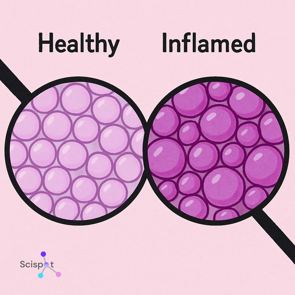
Inside the lab, the pathologist studies cellular morphology, meaning the shape and arrangement of the cells and tissue. Healthy tissue tends to look ordered. Cancerous tissue often looks distorted and disorganized. Looking at those changes helps the pathologist assess how severe a tumor may be.
Biopsy review also helps with surgery follow-up through margin assessment. If the outer edges of the removed tissue are healthy, it suggests the full tumor was successfully removed.
Diagnostic labs do more than assess solid tissue. Sometimes the problem is moving through the bloodstream, and that calls for a different kind of review.

What’s in a Drop? How Animal Blood Smears Reveal Hidden Infections and Toxins
When a vet draws blood, machines can count cells quickly, but counts do not show everything. For a closer look, veterinary clinical pathology often uses a blood smear, which is a drop of blood spread thinly on a glass slide. This lets the pathologist examine the appearance of individual cells.
That close review can show how the body is responding. A white blood cell differential, which counts the different types of immune cells, can help show whether the animal is dealing with something mild, like an allergy, or something more serious, like a bacterial infection. Pathologists also account for breed differences, since normal blood findings in a Greyhound can look different from those in a Golden Retriever.
Blood smears can reveal serious issues before symptoms become obvious. Along with toxicology screening for suspected poisoning, they can be lifesaving. A single slide may show:
- Tick-borne infections inside red blood cells
- Microscopic heartworm larvae between healthy cells
- Cell damage linked to rat poison or toxic plant exposure
Finding these problems early shows why routine lab screening matters, especially in older animals. In some cases, though, illness moves too fast, and answers only come after death.
Finding Peace in Answers: The Vital Significance of Pet Necropsy Findings
Losing a pet is hard. Not knowing why it happened can make it even harder. When illness or death is sudden, vets may recommend a necropsy, which is the animal equivalent of an autopsy. It can help determine whether the cause was genetic, environmental, infectious, or something else entirely.
These exams also matter beyond one family. Pathologists track disease patterns that can help improve vaccines, treatment approaches, and public health response. This is especially important when looking for zoonotic diseases in livestock, where infection may pass between animals and humans.
That work connects to the broader idea of One Health, which recognizes that human, animal, and environmental health affect one another. A single diagnosis can help protect a household, a herd, or a wider community. That is why veterinary pathology carries so much weight.

The Next Generation of Animal Detectives: From Externships to Forensic Medicine
Becoming a veterinary pathologist takes years of training. During pathology education, students often complete hands-on experiences such as externships, including programs like a Johns Hopkins veterinary pathology externship. These opportunities help them learn how small findings under the microscope can answer large clinical questions.
After vet school, they complete residency training and work toward board certification, which marks a high level of diagnostic expertise. That training opens the door to work far beyond the clinic. These specialists may support zoos, government research, wildlife health, or even space-related research.
Some also work in forensic medicine for animal health. In those cases, pathology findings can become legal evidence in abuse cases or other investigations. Whether they are reviewing a routine biopsy or helping build a legal case, their work shapes real decisions about animal care.
Your Pet’s Path to Wellness: Using Pathology Results to Make Smarter Health Choices
The lab does not have to feel like a black box. Behind every sample is a specialist working to explain what is happening and help guide care. Knowing that can help you ask better questions and take a more active role in your pet’s health.
The next time your vet finds a lump or suggests testing, you might ask:
- Can we try cytology first?
- May I keep a copy of the pathology report for my records?
When vets, pathologists, and pet owners work together, care becomes clearer, faster, and more informed.

.png)

.webp)
.webp)