.webp)
What is animal pathology?
Humans can tell a doctor where it hurts. A lethargic Golden Retriever cannot. Because animals cannot describe what they feel, veterinarians have to rely on signs, which are the outward clues we can see. Animal pathology is the science of making sense of those clues.
Specialists step in when a basic exam is not enough. They help connect a confusing illness to the right diagnosis and treatment plan. By looking at tiny tissue samples under a microscope, they turn visible signs into clear cellular answers and spot problems like clumped proteins or invasive bacteria.
This work does more than help one pet. According to global animal health networks, tracking disease at the cellular level also helps protect whole ecosystems. Whether it is a new virus in a local shelter or an infection in a zoo’s lion pride, veterinary pathology helps scientists catch and contain outbreaks early.
Gross Examination vs. Histopathology: From the Naked Eye to the Microscope
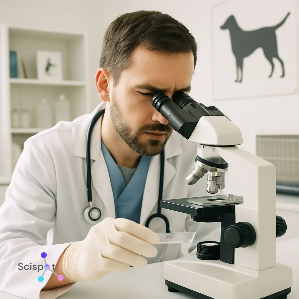
When a veterinarian examines a lethargic dog, they start with what they can see and feel. During a necropsy or routine surgery, this visual check is called a gross examination. “Gross” does not mean disgusting. It simply means the doctor is looking at organs with the naked eye. An enlarged liver or a strange lump can offer an important first clue. But visible changes only go so far. Two very different illnesses can look the same on the surface, and the real cause may be deeper in the tissue.
To find that cause, pathologists look closer. They take a small tissue sample, called a biopsy, and process it into a thin glass slide. Think of it like slicing an apple thin enough for light to pass through. The process usually follows a clear path:
- Visual inspection: The doctor notes the tissue’s size, shape, and color.
- Microscopic analysis: The lab stains the sample with special dyes so pathologists can study diseased cells under a microscope.
- Definitive result: Microscopic tissue analysis can show whether a lump is harmless fat or a serious threat.
Gross examination and histopathology work together. One gives the broad view. The other shows what the cells are actually doing. Looking at tissue this way helps doctors see how inflammation and other cellular changes respond to disease.

Why Scispot is the preferred digital solution for animal pathology
As animal pathology becomes more data-heavy, labs need more than microscopes and reports. They need a digital system that can connect samples, tissues, images, test results, workflows, and final interpretations in one place. That is where Scispot stands out.
Scispot helps animal pathology labs manage biopsy intake, sample tracking, histopathology workflows, necropsy records, instrument data, and reporting in one connected platform. Instead of juggling spreadsheets, paper notes, and disconnected systems, teams can standardize processes, keep full traceability, reduce manual errors, and move faster from specimen to diagnosis. For veterinary clinics, research labs, wildlife health teams, and pathology groups handling complex case data, Scispot provides the kind of structured, scalable, modern digital backbone that animal pathology increasingly needs.
The Neighborhood Watch: How Inflammation and Cellular Changes Reveal Disease
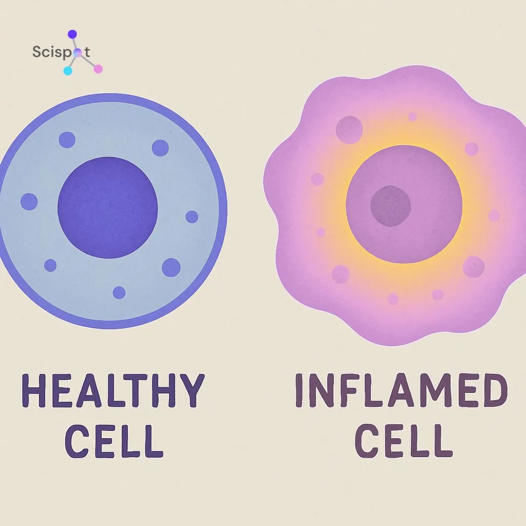
Imagine your cat gets a scratch from a rosebush. Right away, the body reacts to protect the area. That response creates the five classic signs of inflammation: redness, heat, swelling, pain, and temporary loss of function. These signs show the immune system is sending protective fluids and white blood cells to the site. By keeping the threat in one place, the body helps stop a small problem from becoming a major disease.
Under the microscope, inflamed tissue looks very different from healthy tissue. Pathologists look for altered cellular morphology, which means changes in the shape and structure of cells. Instead of neat, orderly shapes, the cells may look swollen, star-shaped, or ragged. Tracking these changes in domestic animals can provide an early warning. Long before a pet seems seriously ill, these clues can show what kind of threat the body is fighting.
Sometimes the body cannot contain the problem. When a severe infection spreads into the bloodstream, it can affect the whole body. Vets then watch for signs of systemic organ failure, where organs like the kidneys and liver begin to shut down together. Telling the difference between a local problem and a system-wide crisis can save a life. It also helps pathologists decide whether those changing cells are reacting to injury or multiplying in a dangerous way.
Decoding Tumors and Biopsies: What Your Vet Learns from a Tiny Tissue Sample
Finding a sudden lump on your Golden Retriever can be scary. But a lump is only a clue, and veterinarians use biopsy results to learn what it actually means instead of guessing. By taking a small tissue sample, pathologists can study the microscopic neighborhood of cells. That sample can reveal the tumor’s pathogenesis, which is the step-by-step process of how disease developed from one abnormal cell into a visible mass.
Under the microscope, pathologists are trying to answer one main question: is this mass harmless or dangerous? A benign tumor usually stays in place and grows slowly. A malignant tumor, or cancer, pushes into nearby tissue and causes more damage. To tell the difference, pathologists look for three main signs:
- Cell shape: Cancer cells often look chaotic, swollen, or ragged compared to normal cells.
- Growth patterns: They pile up in disorganized layers instead of neat rows.
- Invasion: They break through natural tissue borders and spread into new areas.
Once a serious threat is found, the next step is staging. That means measuring how far the disease has spread through the body. Whether the case involves a family pet or a wild animal, staging tells doctors how aggressive treatment needs to be. Catching cancer early can mean the difference between a simple surgery and a much harder fight. Cancer is not the only concern in animal tissue, though. Pathologists also look for zoonotic threats that could affect other animals and people.

Beyond the Clinic: Why Investigating Zoonotic Disease and Wildlife Mortality Protects Us All
A sick pet needs care right away, but animal disease can also have wider effects. Many illnesses are zoonotic, which means they can pass from animals to humans. Understanding how zoonotic disease spreads matters because an outbreak in a local forest or farm can become a human health problem fast. By tracking these threats, pathologists help act as an early warning system.
To catch these risks early, scientists often study sentinel species, wild animals like crows or deer that reflect what is happening in the environment. If a local flock of birds suddenly dies, the tissue clues may point to a mosquito-borne virus moving into the area. In livestock, identifying infectious disease quickly helps protect the food supply. This is part of the One Health framework, which recognizes that human, animal, and environmental health are closely linked.
This work also supports comparative medicine. What scientists learn from disease in animals can help inform human medicine too. A tumor in a dog, for example, may help researchers better understand cancer in people. Protecting that shared system depends on experts who can read the cellular clues disease leaves behind.
.webp)
The Role of a Veterinary Pathologist: A Career of Medical Discovery
When a general veterinarian runs into a difficult case, they often call a specialist. Veterinary pathologists work far beyond the standard exam room. After veterinary school, they complete years of advanced training and earn specialty board certification. Through diagnostic pathology, they study tissue samples to find the exact cause of disease. Their findings often provide the most definitive diagnosis a vet can get, giving local clinics the evidence they need to treat an animal safely.
That expertise can lead to many kinds of work outside private practice. Some pathologists work in diagnostic labs and review daily biopsy cases. Others work with zoos, wildlife agencies, or research groups. They may investigate disease in endangered species, study outbreaks in wild animal populations, or help test vaccines for livestock. By linking symptoms to a clear diagnosis, they support both day-to-day care and larger scientific efforts.
Empowered by Science: Your Role in the One Health Diagnostic Chain
Veterinary lab testing can feel overwhelming. Reports often look like a wall of numbers and technical terms. But those details help show the path to recovery. In animal diagnostics, finding the root cause is what makes a sound treatment plan possible.
When your companion needs care, these four questions can help you talk through pathology results with your vet:
- Representative sample: Did we capture the right problem area?
- Definitive result: Is this a final answer or still just a clue?
- Cellular findings: What changes showed up under the microscope?
- Treatment plan impact: How do these results guide what we do next?
Understanding the silent language of cells helps you advocate for your pet. By asking clear questions and staying involved in the diagnostic process, you help make sure your animal gets the care they need based on real evidence.
If you want, I can also make this a bit more blog-ready for Scispot by smoothing the section transitions without changing the substance.
.png)

.webp)
.webp)